Frog anatomy is built around two facts that no other vertebrate group combines: the body breathes through its skin, and the back legs are long enough to throw the body 20 times its own length in a single jump.
The frog (around 7,500 species in the order Anura) carries no neck, no tail in adults, no ribs that meet a sternum, and a backbone of only 9 free vertebrae plus a single fused rod (the urostyle) that turns the lower back into a built-in catapult.
The skin is a respiratory organ, a salt and water regulator, and a chemical defence wrapped in one. Frogs range from the 7.7 mm Paedophryne amauensis of New Guinea, the smallest vertebrate alive, to the 32 cm goliath frog (Conraua goliath) of West Africa at over 3 kg, but every species shares the same short body, long hindlimbs, paired bulging eyes, and porous moist skin that defines the group.
Frogs sit in the order Anura (“without tail”) inside the class Amphibia. The order splits into around 55 families, of which true frogs (Ranidae), tree frogs (Hylidae), poison frogs (Dendrobatidae), and the toads (Bufonidae) are the most familiar. The line between “frog” and “toad” is not a strict taxonomic one: toads are a subset of frogs with thicker drier skin, shorter legs, and more time spent on land, but they share the same body plan. Most frogs hatch from eggs into tadpoles with gills and a tail, then metamorphose into the adult body over weeks to years, depending on species.
The adult body covered on this page is the result of that transformation. Several existing AnimalCorner pages cover individual systems in detail: external anatomy, skeleton, muscles, mouth cavity, brain, eye, and heart. The sections below give the integrated full-body view.
Frog Body Plan: No Neck, No Tail, No Sternum
A frog’s body is short, broad, and wedge-shaped, optimised for the launch of a jump rather than the steady-state walk of most vertebrates. Three structural simplifications drive the shape. The skull connects to the spine at a single rounded joint that allows up-and-down nodding but no real side-to-side rotation, so the frog has no functional neck. The tail present in the tadpole is reabsorbed during metamorphosis and disappears in the adult; the only trace is the urostyle, a single rod-shaped bone formed by the fusion of the original tail vertebrae.

The ribs that exist in most other vertebrates are reduced or absent in frogs. The body wall is held in shape by the skin, the abdominal muscles, and the connection between the pectoral girdle and the pelvic girdle through the body cavity itself. The result is an open body cavity with the heart and lungs at the front, the liver and stomach in the middle, and the kidneys, intestines, and reproductive organs at the back.
The body is built around four limbs. The front legs are short, with four toes that lack webbing in most species, used to brace the landing after a jump and to push the head up off the ground at rest. The back legs are several times longer than the front legs, with five toes, full webbing in aquatic species, and a folded posture at rest that hides their full extension. When the back legs straighten in the powerstroke of a jump, the bullfrog (Lithobates catesbeianus) launches its 500 g body up to 2 m forward and 1 m up. The longest jumper in proportion to body length, the South African sharp-nosed frog (Ptychadena oxyrhynchus), throws its 4 cm body over 4 m in a single jump, more than 100 times its own length. The diagram below labels the visible body parts.
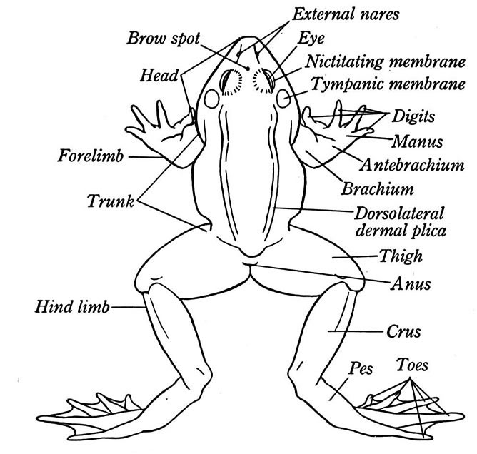
The Frog Skeleton: 9 Vertebrae, the Urostyle, and the Jumping Pelvis
A frog has the simplest spine of any tetrapod. The whole vertebral column carries 9 free vertebrae plus the urostyle: 1 cervical vertebra (the atlas, which connects to the skull), 7 trunk vertebrae (which carry small lateral processes but no full ribs in most species), the sacral vertebra (which articulates with the pelvis), and the urostyle. Compare this to the 33 vertebrae in a human spine. The reduction is the result of fusion and loss during the transition to a jumping body plan. The urostyle and the sacrum together form a rigid rear platform against which the long muscles of the back and pelvis can pull during the launch of a jump.
The pelvis is unusual. The two ilia (the upper bones of the pelvis) are stretched forward into long parallel rods that run along the underside of the trunk vertebrae and join the urostyle at a movable joint. The whole pelvis can rock fore-and-aft against the spine, adding extension distance to the leg drive at the moment of takeoff. The front limbs carry the standard tetrapod bones (humerus, radius, ulna, carpals, metacarpals, phalanges), with the radius and ulna fused into a single radio-ulna in adults. The back limbs carry a long femur, a fused tibia and fibula (the tibiofibula), an extended ankle (the astragalus and calcaneum, which form a second long-bone segment), and the foot. The fused leg bones add length without adding the joint count that would lose energy on the way through the powerstroke. For a full diagram of the bones, see the Frog Skeleton Anatomy.
Frog Skin: Breathing, Drinking, Defending
A frog’s skin is a working organ, not a passive cover. It is thin (around 0.1 to 1 mm in most species), kept moist by mucous glands, and crossed by a dense capillary network that carries blood to within 25 micrometres of the outer surface. Oxygen and carbon dioxide diffuse through the skin in both directions. In a resting frog at cool temperatures, around 50 percent of oxygen uptake and almost all of carbon dioxide release happens through the skin rather than the lungs. In a hibernating frog at the bottom of a winter pond, the lungs shut down and 100 percent of gas exchange is cutaneous. Some species (the lungless salamanders of the family Plethodontidae are not frogs but illustrate the same point) have lost their lungs altogether and live on skin breathing alone.
A frog drinks through its skin, not through its mouth. A patch on the underside of the body and the inner thighs (the “drinking patch” or pelvic patch) absorbs water from the ground or from a damp surface by osmotic uptake, which the frog regulates by changing the permeability of the skin under hormonal control. A frog can replace 30 percent of its body water in a single hour through the patch. The same skin loses water in dry conditions, which is why most frogs need to stay near water, although the waterproof tree frogs (Hylidae) and waxy monkey frogs (Phyllomedusa) have evolved waxy lipid coatings on the skin that cut water loss to 1 to 5 percent of the rate in a typical frog and let them survive in arid conditions.
Frog skin is a chemical defence as well. Mucous glands on the skin secrete antibacterial and antifungal peptides that protect against the pathogens that thrive in the moist environment of the skin surface. Granular glands deeper in the skin produce alkaloid toxins in many species, secreted under stress. The poison dart frogs of Central and South America (Dendrobatidae) carry the most lethal of these: a single golden poison frog (Phyllobates terribilis) carries enough batrachotoxin in its skin to kill 10 to 20 humans. The frog does not produce the toxin from scratch; it takes the alkaloids from the ants and beetles in its diet and re-secretes them through the skin. Frogs raised in captivity on a different diet are not toxic.
The skin sheds in one piece, every few days to every few weeks depending on species and temperature. The frog wriggles out of the old skin from the back forward, shrugs the loose skin over its head, pulls it free with the front feet, and then swallows it. The shed skin contains useful protein and the frog reabsorbs it through the digestive tract. The new skin underneath is brighter and more permeable.
Eyes That Push Food Down the Throat
A frog’s eyes bulge out from the top of the skull because they double as part of the swallowing mechanism. When the frog closes its eyelids, the eyeballs sink down into the mouth cavity through openings in the roof of the skull and press against the food on the tongue, pushing the prey item back into the throat. A frog that swallows a large insect is visible blinking with each swallow as the eyes drive the prey down. The eyes are large in relation to the head: a bullfrog eye is around 7 mm across on a 5 cm skull, with a pupil that closes to a horizontal slit in some species or stays round in others.

Frog vision is built for movement detection at close range rather than acuity at distance. The retina contains four types of cones, a wide range of rods, and a layer of specialised retinal ganglion cells called “bug detectors” that fire only when a small dark moving object crosses the visual field at the right speed. A frog will starve in front of a pile of dead flies because the prey is not moving. Frogs see colour into the green and blue range and have the rare ability to see colour at light levels too dim for human cones, by repurposing the rod cells for colour discrimination.
Each eye is protected by an upper eyelid, a lower eyelid, and a transparent third eyelid (the nictitating membrane) that sweeps across the eye underwater or during wrestling matches. For more on the eye, see Frog Eye Anatomy.
Mouth, Tongue, and the Sticky-Tongue Strike
A frog’s mouth opens wider than its skull is long. The lower jaw drops and the upper jaw lifts at the same time, hinging at a joint at the back of the skull. Most frogs carry small recurved teeth around the rim of the upper jaw (the maxillary teeth) and on the roof of the mouth (the vomerine teeth on the vomer bone), used to hold prey rather than to chew. The lower jaw carries no teeth in most species. The toads (Bufonidae) and the South American horned frogs (Ceratophrys) have lost the maxillary teeth altogether; the gastric brooding frog of Australia (now extinct) was unique in carrying teeth in the lower jaw.
The tongue is attached to the front of the lower jaw rather than the back of the mouth, the opposite of the mammalian arrangement. At rest the tongue lies folded forward on the floor of the mouth with its free end pointing backward into the throat. When the frog strikes at prey, the tongue flips out of the mouth like a coiled spring released, hits the prey at speeds up to 4 m/s, sticks to it through a combination of suction and a coating of dense saliva that turns from a watery liquid to a sticky paste under the impact force, then flips back into the mouth carrying the prey. The whole strike takes around 0.07 seconds. The frog’s saliva is around 50,000 times more viscous than human saliva at the moment of impact and around the viscosity of honey. After the strike, the saliva returns to a watery state and the prey slides into the throat. For more on the inside of the mouth, see Frog Mouth Anatomy.
Three-Chambered Heart and a Lung that Shares Work with the Skin
A frog has a three-chambered heart: two atria (left and right) feeding a single ventricle. Oxygen-rich blood from the lungs returns to the left atrium; oxygen-poor blood from the body returns to the right atrium; both atria empty into the same ventricle, which pumps the mixed blood out to the body and the lungs. In a true mammalian or bird heart the two streams stay separate through a divided ventricle. In the frog they mix, but the mixing is less complete than the structure suggests: spiral folds inside the ventricle and a partial septum direct most of the oxygen-rich blood toward the body and most of the oxygen-poor blood toward the lungs and skin. The arrangement is a working compromise that suits an amphibian whose oxygen demand changes with body temperature, activity, and depth underwater. For more on the heart, see Frog Heart Anatomy.
The lungs are paired sacs that sit on each side of the heart, simpler than mammalian lungs and without a diaphragm to drive them. A frog breathes by buccal pumping rather than by chest expansion. The frog lowers the floor of the mouth to draw air in through the nostrils, closes the nostrils, then raises the floor of the mouth to push the air down the throat into the lungs. To exhale, the body wall contracts on the lung and the air is pushed back out. The whole cycle is visible from outside the frog as the throat pulses and the body sides flex. At rest at cool temperatures, around 30 to 50 percent of oxygen uptake is through the lungs and the rest is through the skin. During heavy exercise or warm conditions, the lungs take on more of the load. During hibernation underwater, the lungs shut down and the skin handles 100 percent of gas exchange.
Digestive and Excretory Systems
The frog’s digestive tract is short by mammalian standards, around 2 to 3 times the body length. The mouth opens into the oesophagus, which drops into a J-shaped stomach. The stomach secretes hydrochloric acid (pH around 2 in most frogs) and pepsin to begin protein digestion. The small intestine runs from the stomach through a series of loops in the body cavity and connects to the short large intestine, which empties into the cloaca. The liver is large and lobed, the largest single organ in the body cavity, and produces bile that the gallbladder stores and releases through the bile duct into the small intestine. The pancreas sits in a sheet of tissue between the stomach and the duodenum and produces digestive enzymes and the hormones insulin and glucagon. Frogs swallow prey whole and digest it slowly: a bullfrog that takes a large insect can spend a day or two processing the meal.
The kidneys are paired flat structures running along the back of the body cavity, dorsal to the digestive tract. They filter blood and produce urine that drains down the ureters into the cloaca and then into the urinary bladder, an extension of the cloaca that can hold up to 30 percent of the frog’s body mass in stored water. The bladder doubles as a water reserve: a frog in a dry burrow can reabsorb water from the bladder back through the bladder wall into the bloodstream during drought. The cloaca handles digestive waste, urinary waste, and reproductive output through a single opening, the same arrangement as in birds, reptiles, and most other amphibians.
Nervous System and Vocalisation
The frog brain is small (around 0.05 to 0.1 g in a common frog) and divided into the same regions as other vertebrate brains: the forebrain (olfactory bulbs and cerebral hemispheres), the midbrain (with prominent optic lobes that process the visual signal from the bug-detector cells in the retina), the cerebellum (small in frogs, since the body does not need fine-tuned balance), and the hindbrain (the medulla oblongata, controlling breathing and heart rate). The spinal cord runs from the brain through the vertebral column and ends short of the urostyle, with peripheral nerves branching out to the limbs and body wall. For more, see Frog Brain Anatomy.
The vocal apparatus sits in the throat. The larynx contains paired vocal cords that vibrate as air passes from the lungs across them. Most male frogs carry one or two vocal sacs, expandable pouches of skin under the throat or at the corners of the jaws that act as resonance chambers and amplify the call. The coqui frog of Puerto Rico (Eleutherodactylus coqui) produces a 100 dB call at 1 m, the loudest call of any frog and louder than a chainsaw. Calls are species-specific and serve as the main mate recognition signal. Females hear through a tympanum (eardrum) on each side of the head, set behind the eye, and through the inner ear, which is wired to the lung wall through a sound-conduction pathway that lets the frog hear airborne sound during a deep underwater dive without exposing the eardrum to water.
Reproductive Anatomy and the Tadpole-to-Adult Transition
Frogs reproduce by external fertilisation in around 90 percent of species. The male grips the female from above in an embrace called amplexus, often for hours and sometimes for days, and releases sperm over the eggs as the female releases them into the water. The eggs hatch into tadpoles within days to weeks. A tadpole carries a long tail, paired internal gills, a single round mouth with rasping keratin tooth-rows, a long coiled gut for a herbivorous diet, and no limbs. Over weeks to years (depending on species and temperature), the tadpole metamorphoses: the back legs grow first, then the front legs (hidden under the gill covers until they emerge), the tail shortens and is reabsorbed, the gills are replaced by lungs, the mouth widens and grows the adult jaw structure, the gut shortens for a carnivorous diet, and the eyes migrate to the top of the head.
Some frogs skip the tadpole stage altogether. Direct-developing frogs (most of the family Eleutherodactylidae and many Microhylidae) lay eggs that hatch as tiny adults; the tadpole development happens inside the egg. Other frogs go in the opposite direction and never metamorphose: the axolotl is not a frog (it is a salamander) but illustrates the same retention of the larval body plan into adulthood. The full life cycle is covered in detail at Life Cycle of a Frog.
Frog vs Toad vs Salamander: Anatomy Comparison
“Amphibian” covers three living groups that share a thin permeable skin and a life history that includes water in some form, but their adult body plans differ across the skeleton, the limbs, and the skin.
| Feature | Frog (true frog, e.g. Ranidae) | Toad (Bufonidae) | Salamander or Newt (Caudata) |
|---|---|---|---|
| Tail in adult | None | None | Present, full length |
| Skin | Smooth, moist | Dry, warty, thicker | Smooth, moist |
| Hindlimbs | Long, built for jumping | Shorter, built for walking and short hops | Same length as forelimbs; built for walking |
| Eyes | Large, bulging | Smaller, less prominent | Small, set on the side of the head |
| Vertebrae | 9 free + urostyle | 9 free + urostyle | 10 to 60+ vertebrae plus a tail of additional vertebrae |
| Teeth | Small maxillary and vomerine teeth (none in lower jaw) | None (or vomerine only) | Small teeth in both jaws in most species |
| Habitat preference | Near water through life | Damp land, returns to water to breed | Cool damp ground, water or land depending on species |
| Order | Anura | Anura (a subset of frogs) | Caudata (Urodela) |
For deeper coverage of individual systems and related amphibians, see the AnimalCorner pages on External Anatomy of a Frog, Frog Skeleton Anatomy, Frog Muscles Anatomy, Frog Mouth Anatomy, Frog Brain Anatomy, Frog Eye Anatomy, and Frog Heart Anatomy. For the broader amphibian group, see the Amphibian hub, Newt Anatomy, Salamander Skeletal Anatomy, and the State Amphibians of the USA. Browse the full Animal Anatomy hub for comparable species.